
No products in the cart.
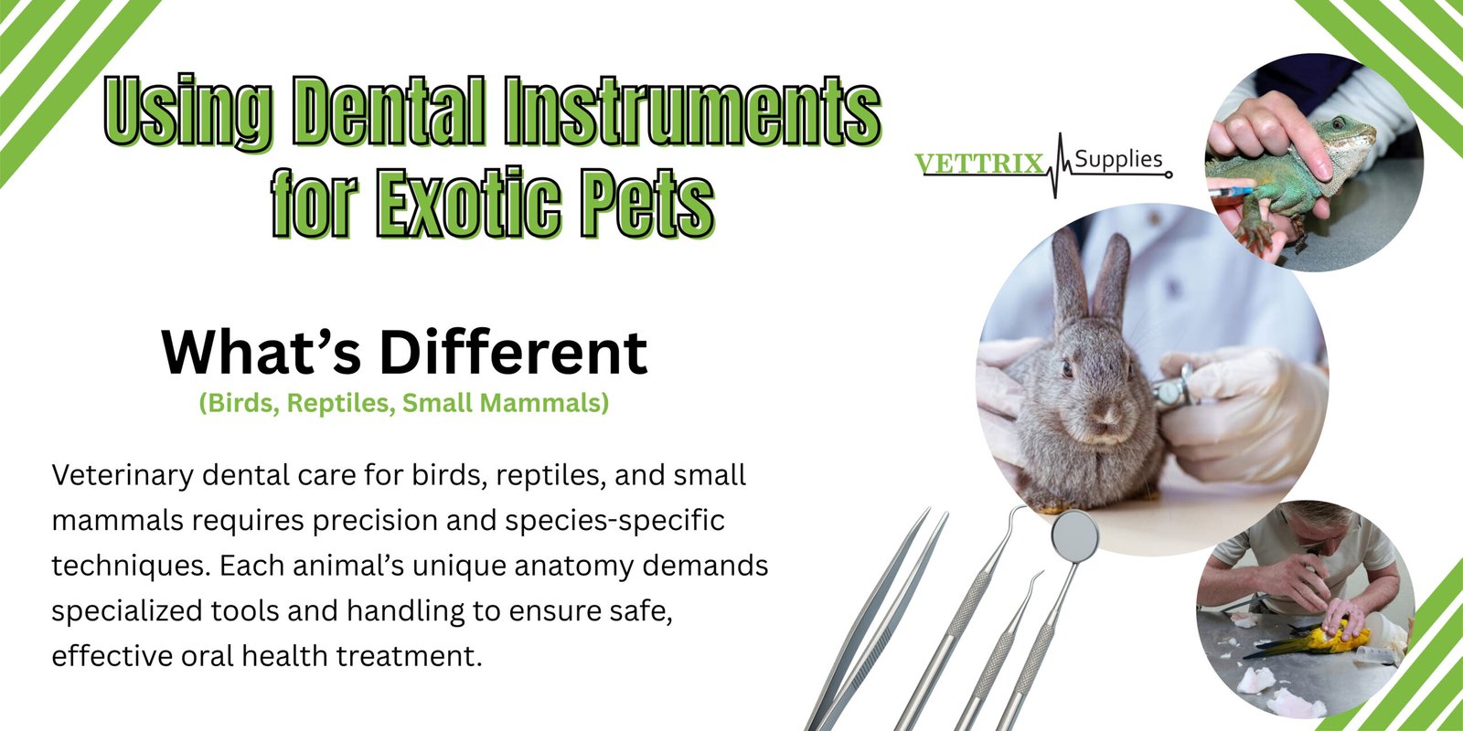
Using Dental Instruments for Exotic Pets: What’s Different (Birds, Reptiles, Small Mammals)
Mastering Exotic Pet Dental Care: Essential Instruments for Birds, Reptiles, and Small Mammals
Caring for the unique dental needs of exotic pets requires a specialized toolkit designed to accommodate their diverse oral structures. This ensures safe and effective examination, diagnosis, and treatment for birds, reptiles, and small mammals. Without the right instruments, common issues like overgrown incisors in rabbits or beak malocclusion in parrots can lead to significant pain, malnutrition, and persistent infections. This guide will explore how distinct tooth and beak anatomies dictate instrument selection, highlight essential tools for each animal group, and detail best practices for their application, maintenance, and sterilization to achieve optimal patient outcomes. You’ll discover:
- The key differences in dental anatomy across small mammals, reptiles, and birds
- Critical diagnostic, treatment, and surgical instruments vital for exotic dentistry
- Specific instrument applications for small mammal, reptile, and avian dental procedures
- Essential anesthesia protocols, pain management strategies, and post-operative care
- Proven methods for instrument upkeep and sterilization
By the end of this guide, veterinary professionals and dedicated pet owners will gain a comprehensive understanding of why precision instruments are indispensable and how each tool contributes to safe, effective dental care for exotic companions.
Unpacking the Unique Dental Anatomies of Exotic Pets
The distinct variations in tooth and beak structures among exotic pets fundamentally shape instrument selection, enabling precise examination and treatment. A thorough understanding of these differences is crucial for preventing procedural complications and implementing targeted oral health strategies. Let’s compare the core dental structures of rabbits, reptiles, and birds, and consider their implications for instrumentation and technique.
| Species | Dental Structure | Maintenance Implication |
|---|---|---|
| Rabbits & Rodents | Continuously growing (hypsodont) teeth | Requires regular trimming, floating, and rasping |
| Reptiles | Acrodont, pleurodont, or thecodont teeth | Extraction tools vary based on attachment type |
| Birds | Keratin-covered beak instead of teeth | Beak files and rotary tools replace tooth cutters |
Each unique morphology necessitates specialized approaches: the continuous growth of rodent teeth can lead to spurs and malocclusion; reptile tooth attachment dictates the force and angle of extraction; and avian keratin requires shaping rather than extraction. These fundamental distinctions guide the selection of examination, diagnostic, and treatment instruments tailored for each group.
How Do Small Mammal Teeth Differ from Other Species?

Small mammal teeth are characterized by continuous growth, particularly their open-rooted incisors and molars—a condition known as hypsodonty. This adaptation demands frequent occlusal adjustments to prevent overgrowth, which can result in malocclusion, painful buccal ulcers, and significant weight loss. Molar rasps and incisor cutters must be designed to navigate narrow oral cavities while applying controlled abrasion or cutting force.
Hypsodont teeth necessitate instruments with angled shafts and extended handles for optimal reach. Cheek dilators are essential for retracting soft tissues and improving access, while magnification aids in visualizing the occlusal surfaces. The management of constantly erupting teeth clearly distinguishes small mammal dentistry from the care required for reptiles or birds, whose teeth are fixed or absent.
What Are the Distinctive Dental Features of Reptiles?
Reptile dentition typically falls into three primary attachment types: acrodont (tooth fused directly to the jawbone), pleurodont (tooth attached to the inner surface of the jaw), and thecodont (tooth set within bony sockets). Each type influences the torque required for extraction and the design of the instruments used. Acrodont extractions often require precision chisels, while pleurodont teeth respond best to angled luxators. Thecodont tooth removal techniques are more akin to mammalian forceps procedures.
Reptile oral cavities are frequently affected by stomatitis and mouth rot, necessitating instruments that can reach deep oral sulci without causing further damage to delicate mucosa. Debridement curettes and small scalers must be carefully selected to match the species-specific jaw shape. Recognizing the tooth attachment type is paramount for optimizing instrument selection and minimizing the risk of jaw fracture.
Why Do Birds Require Beak-Specific Dental Care Instead of Teeth?
Birds possess keratin-covered beaks, not true teeth, and overgrowth or misalignment can severely impair their ability to feed, preen, and vocalize. Beak anatomy consists of a bony core covered by a keratin sheath. are used to reshape the keratin sheath without damaging the underlying vascular bone. must be designed to create smooth contours and provide precise abrasion.
Beak care utilizes appropriately sized for species ranging from parakeets to macaws, each requiring different diameters and grit levels. Accurate reshaping is crucial for maintaining proper alignment and wear patterns influenced by diet. Understanding avian beak biomechanics underscores the need for specialized trimming tools rather than traditional dental burs or forceps.
Which Dental Instruments Are Essential for Exotic Pet Dentistry?
The for exotic pets span examination, diagnostics, treatment, and surgery, all calibrated for varying mouth sizes and anatomies. Selecting the appropriate set enhances precision, reduces procedural time, and significantly improves animal welfare. Here is a categorized list of core devices indispensable for any exotic veterinary dental kit.
- Mouth Gags and Cheek Dilators for safe and secure oral cavity access
- Dental Mirrors and Tongue Depressors for visualizing hard-to-reach surfaces
- Intraoral Radiography Sensors and Portable CT Units for detailed diagnostic imaging
- Luxators, Elevators, and Extraction Forceps for atraumatic tooth removal
- Dental Burs, Molar Cutters, and Rasps for precise occlusal adjustments
- Ultrasonic Scalers and Debridement Curettes for effective biofilm and lesion management
Each instrument must be available in a variety of sizes and angles to accommodate the diverse needs of species, from the smallest rodents to the largest parrots. A comprehensive inventory is key to supporting both routine preventive exams and complex surgical interventions with minimal risk.
What Examination and Diagnostic Tools Are Used for Exotic Pets?
Oral examinations in exotic pets require adapted devices that ensure both safety and optimal visibility within small or narrow mouths. with adjustable locking mechanisms maintain jaw opening without causing tissue trauma, while gently retract soft tissues laterally. with angled heads and integrated fiber-optic lighting are crucial for revealing occlusal and lingual surfaces. Advanced , including micro-size intraoral sensors and portable CT scanners, provides high-resolution images essential for diagnosing tooth roots, jaw fractures, and abscesses.
significantly enhance diagnostic precision by improving visualization of fine details, aiding in the detection of subtle spurs in rabbits or early-stage stomatitis in reptiles. Collectively, these tools form the bedrock of a proactive, evidence-based approach to exotic dental evaluations.
What Treatment and Surgical Instruments Are Specialized for Exotic Animals?
The treatment and surgical phases of exotic pet dentistry rely on meticulously engineered for precision within confined spaces. with fine, curved tips are designed to gently separate periodontal ligaments in pleurodont reptiles and hypsodont rodents. are used to transfer force from luxation sites, minimizing bone trauma. are ideal for navigating the narrow orbits found in ferrets and bearded dragons. , both carbide and diamond, are available in extended, slender designs for contouring molars and adjusting occlusal planes. are employed to sever overgrown teeth, while are used to create smooth enamel surfaces. equipped with fine-tip inserts effectively remove plaque and calculus with minimal vibration.
are utilized for precise wound closure following jaw fracture repairs or tooth extraction sites, promoting rapid healing.
How Do Instrument Designs Vary Between Birds, Reptiles, and Small Mammals?
| Instrument Type | Species Adaptation | Primary Purpose |
|---|---|---|
| Luxator | Curved tip specifically for pleurodont teeth (reptiles) | Gentle severing of periodontal ligaments |
| Molar Rasp | Angled head with fine grit for rodent molars | Controlled smoothing of molar surfaces |
| Beak File | Flat, broad rasp designed for avian beaks | Reshaping and smoothing the keratin sheath |
| Extraction Forceps | S-shaped shaft for narrow oral cavities (ferrets, lizards) | Atraumatic tooth removal |
| Ultrasonic Scaler Tip | Reduced diameter insert for small mouths | Effective removal of biofilm and calculus |
These specialized adaptations ensure that function efficiently and safely, without causing harm to adjacent tissues. Matching instrument design to the specific anatomy of the patient optimizes treatment outcomes and significantly reduces the likelihood of procedural complications.
How Are Dental Instruments Applied in Small Mammal Dentistry?
Small mammal dental care involves a combination of precise trimming, abscess management, and occlusal correction, utilizing specialized to address the challenges posed by continually erupting teeth. Procedures typically begin with ensuring atraumatic access, followed by targeted treatment, and concluding with preventive maintenance to sustain optimal oral health.
Initially, a and are employed to stabilize the oral cavity, providing clear visibility of both incisors and molars. are used for trimming overlong front teeth, while and are employed to level molar occlusal surfaces. and assist in the extraction of fractured or maloccluded teeth. Following treatment, antiseptic irrigation and appropriate suture placement support healing and minimize the risk of infection.
What Tools Are Used for Rabbit and Rodent Incisor and Molar Care?
- Incisor Cutters: Available in scissor-style or guillotine blade designs for clean, precise incisor trims.
- Molar Rasps: Handheld or motorized files designed to gently abrade molar spurs and smooth rough surfaces.
- Cheek Dilators: Used to gently expand the buccal cavity, improving access without overstretching soft tissues.
These devices work synergistically to correct overgrowth, remove enamel spurs, and restore proper occlusion. Incorporating magnification and angled burs further enhances precision, especially in tight oral spaces.
How Are Dental Abscesses and Malocclusion Treated in Small Mammals?
The treatment of dental abscesses and malocclusion in small mammals integrates -assisted debridement and luxation techniques. Once abscess tracts are identified via radiography, are used to gently separate infected teeth from the surrounding alveolar bone. meticulously remove purulent material, and antimicrobial irrigants are employed to flush out debris. and are crucial for extracting the roots of nonviable teeth, thereby resolving the infection. For malocclusion cases, are used to reshape occlusal surfaces and correct bite planes, while spacers or acrylic caps may be applied to protect treated areas during the healing process. This comprehensive approach effectively addresses both the source of infection and functional alignment, promoting sustained oral health.
What Are Best Practices for Instrument Use in Ferret and Hedgehog Dentistry?
Dental procedures on ferrets and hedgehogs require and protect delicate oral mucosa. facilitate atraumatic removal of fractured canine roots in ferrets. In hedgehogs, are used to adjust molar facets within their restricted oral chambers. It is crucial to always begin with gentle tissue retraction and maintain continuous irrigation to prevent thermal damage. ensure precise closure of extraction sites. Adhering to ergonomic handling principles and conducting regular instrument inspections are vital for consistent performance and patient safety.
What Are the Specialized Dental Instrument Techniques for Reptiles?
Reptile dentistry primarily focuses on managing stomatitis, performing tooth extractions based on specific attachment types, and stabilizing jaw fractures. Successful procedures depend on selecting compatible with acrodont, pleurodont, or thecodont dentition and employing techniques that respect the unique bone density and mucosal fragility of reptiles.
Initial debridement of affected areas utilizes to remove necrotic tissue in stomatitis cases. are employed to facilitate the extraction of acrodont teeth that are fused to the jaw. For pleurodont extractions, are used to detach the tooth along its lingual surface. In thecodont species, can effectively mimic mammalian extraction techniques. Jaw fracture repair involves the use of , all guided by preoperative radiographic imaging.
How Are Stomatitis and Mouth Rot Managed Using Dental Instruments?
The management of stomatitis and mouth rot begins with thorough debridement of inflamed tissue, utilizing along with antimicrobial irrigation. After removing biofilm and necrotic debris, cotton-tipped applicators soaked in topical antimicrobials are used to reach deep sulci. may be employed to gently elevate fibrinous plaques without causing injury to the underlying bone. Treatment concludes with essential dietary and environmental modifications to support mucosal healing and prevent recurrence.
What Instruments Are Used for Tooth Extraction in Acrodont and Pleurodont Reptiles?
Tooth extraction in acrodont reptiles necessitates the use of to carefully detach the tooth from its fused bone structure, thereby minimizing fracture risk. Pleurodont extractions typically involve inserted along the lingual surface to sever the periodontal attachments. are then used to grasp the tooth crown once it has been mobilized. Employing atraumatic techniques is crucial for preserving jaw integrity and reducing postoperative complications.
How Is Jaw Fracture Repair Supported by Dental Tools in Reptiles?
Reptile jaw fractures are stabilized using . Following fracture reduction, are secured in place with to maintain proper alignment. are used to precisely shape and position the plates. Intraoperative radiography is essential for confirming correct placement. After stabilization, and are used to clear surgical debris before soft tissue closure, facilitating rapid bone healing and the restoration of normal feeding function.
How Are Avian Beak Trimming and Oral Examinations Performed?

Avian beak care involves a combination of shaping, examination, and safe handling practices that take into account keratin biomechanics and bird behavior. Proper application is key to achieving smooth beak contours and preventing damage to the vascular core.
The examination process begins with gentle beak opening, assisted by custom beak gags or towel restraint for smaller species. Visual inspection utilizes fiber-optic headlights and magnification to identify any fissures or lesions. Beak trimming employs for minor adjustments and for rapid reshaping. Post-procedural smoothing is achieved using to restore functional ridges essential for feeding and preening.
What Are the Common Beak Trimming Tools for Birds?
- Flat and half-round rasp files, appropriately sized for different beak curvatures.
- Rotary dremels fitted with diamond or carbide burr attachments for efficient keratin removal.
- Fine-grit sanding discs for achieving a smooth, finished surface.
These allow for incremental keratin removal, ensuring the preservation of the underlying bone and vascular tissue. Consistent maintenance is crucial for preventing malocclusion and encouraging natural wear patterns.
How Is Beak Overgrowth Diagnosed and Treated with Specialized Instruments?
Diagnosing beak overgrowth involves comparing the lengths of the upper and lower beaks and identifying any difficulties the bird experiences with feeding. A caliper is used to measure the extent of keratin extension beyond normal anatomical limits. Treatment typically involves . Performing these adjustments in careful increments is essential to prevent thermal damage. Completing the procedure with fine-grit sanding restores the natural beak contours and maintains functional alignment.
What Safety Considerations Are Important When Using Dental Instruments on Birds?
Ensuring safety during avian dental procedures requires a focus on preventing stress and minimizing tissue trauma. Utilize appropriate restraint methods—such as soft towels or custom gags—applied firmly but without excessive force. Maintain continuous cooling when operating rotary tools to prevent heat-induced necrosis. Closely monitor the bird’s respiration, as manipulation of the beak can potentially affect airflow. Always conclude the procedure with antiseptic application to prevent infection and promote keratin regeneration.
What Are the Anesthesia and Pain Management Considerations for Exotic Pet Dental Procedures?
Anesthesia and pain management in exotic pet dentistry necessitate species-specific protocols that effectively balance immobilization, analgesia, and rapid recovery. The choice of can influence the required depth of anesthesia, as certain procedures, like , may demand deeper anesthetic planes to prevent patient movement that could lead to oral tissue injury.
How Does Anesthesia Protocol Vary by Species During Dental Treatments?
Anesthesia protocols are adapted based on the species’ metabolic rate and airway anatomy. Small mammals often receive intramuscular premedication followed by mask induction, while reptiles may require temperature-controlled environments to facilitate the metabolism of inhalant anesthetics. Birds necessitate careful induction via mask, with minimal dead space to prevent hypoxia. Dosages and anesthetic agent choices are tailored to species-specific pharmacokinetics and physiological sensitivities, ensuring adequate muscle relaxation for instrument use without compromising vital functions.
What Pain Management Strategies Support Post-Procedure Recovery?
Post-procedure pain management typically combines local anesthetic nerve blocks with systemic analgesics. Non-steroidal anti-inflammatory drugs (NSAIDs) help reduce inflammation, while opioids, such as buprenorphine, provide potent pain relief for small mammals and reptiles. Transdermal patches or sustained-release formulations can offer continuous analgesia, thereby reducing handling stress. Nutritional support with soft, palatable diets and environmental enrichment further promotes comfort and encourages normal feeding behavior.
How Does Instrument Choice Affect Anesthesia and Procedure Safety?
Selecting that generate minimal vibration and heat can significantly reduce anesthesia time by accelerating procedures and preventing tissue damage. , for instance, produce very little heat, decreasing the need for deep anesthetic planes. allow for faster extractions with less patient movement. Incorporating local nerve blocks can decrease the overall requirement for inhalant anesthetics, promoting faster recovery and lowering anesthetic risk.
How Should Dental Instruments Be Maintained and Sterilized for Exotic Pet Care?
Proper maintenance and of veterinary dental are paramount for ensuring patient safety, prolonging tool longevity, and preventing cross-contamination. must undergo rigorous cleaning, disinfection, and sterilization protocols that align with veterinary best practices and manufacturer guidelines.
What Are Best Practices for Cleaning and Sterilizing Veterinary Dental Tools?
Following each use, should be rinsed thoroughly under warm running water to remove any organic debris. are recommended for use in to effectively dislodge residual material. After rinsing, must be inspected for any signs of damage. Pack in appropriate and process them in an , adhering to the recommended temperature and cycle times. Store sterilized in a dry, closed cabinet to maintain their sterility until their next use.
How Does Proper Instrument Maintenance Improve Treatment Outcomes?
Consistent maintenance preserves sharp edges, prevents corrosion, and ensures accurate force transmission during procedures. Well-maintained burs and cutters require less applied pressure, which in turn reduces tissue trauma and shortens procedural time. Reliable performance leads to more predictable outcomes, minimizes complications, and reinforces practitioner confidence.
What Post-Procedure Care Supports Oral Health in Exotic Pets?
Post-procedure oral health relies on tailored dietary and hygiene recommendations specific to each species. Small mammals benefit from high-fiber diets that encourage natural tooth wear, while reptiles require calcium-rich diets to support bone healing. Birds benefit from access to cuttlebone or mineral blocks to maintain beak health. Regular at-home observation by owners and periodic professional dental examinations by a veterinarian complete a comprehensive oral care plan.
The combination of and meticulous protocols is essential for maintaining the delicate balance of oral health in exotic pets, empowering clinicians to provide precise and compassionate care.
Frequently Asked Questions About Exotic Pet Dental Care
Why is specialized dental care crucial for exotic pets?
Exotic pets have diverse and unique oral structures, unlike traditional companion animals. Without and care, they can suffer from issues like overgrown teeth or beak malocclusion, leading to severe pain, malnutrition, and infections. Tailored care ensures safe and effective examination, diagnosis, and treatment.
How do small mammal teeth differ from those of other species?
Small mammals, such as rabbits and rodents, possess continuously growing (hypsodont) teeth, particularly their incisors and molars. This unique feature necessitates regular trimming, floating, and rasping using and to prevent overgrowth and malocclusion.
What are the main types of reptile dentition?
Reptile dentition typically falls into three categories: acrodont (teeth fused directly to the jawbone), pleurodont (teeth attached to the inner surface of the jaw), and thecodont (teeth set within bony sockets). The type of attachment dictates the specific and techniques required.
Why do birds require beak-specific care instead of teeth?
Birds do not have true teeth; instead, they have keratin-covered beaks. Overgrowth or misalignment of the beak can severely impact their ability to feed and preen. are used to reshape the keratin sheath without damaging the underlying vascular bone.
What essential are needed for exotic pets?
Essential instruments include for access, for visualization, for diagnostics, for extractions, for occlusal adjustments, and for cleaning. These tools must be available in various sizes and angles to suit different species.
How are veterinary maintained and sterilized?
After each use, instruments should be rinsed, cleaned with in an ultrasonic cleaner, inspected for damage, and then packaged in sterilization pouches. They are then processed in an autoclave according to recommended cycles and stored in a dry, closed cabinet to maintain sterility.
Conclusion
Exotic pet dentistry is a specialized field that demands a deep understanding of diverse oral anatomies and pathologies across species like birds, reptiles, and small mammals. The cornerstone of successful treatment lies in the selection and precise application of , meticulously designed to navigate the unique challenges presented by continuously growing teeth, varied tooth attachment types, and keratin-covered beaks.
From diagnostic tools like and to treatment instruments such as , , and , each tool plays a critical role in ensuring safe and effective care. Furthermore, rigorous adherence to species-specific anesthesia protocols, comprehensive pain management strategies, and stringent are indispensable for minimizing risks and optimizing patient outcomes.
Ultimately, consistent and tailored post-procedure care are vital for promoting long-term oral health and enhancing the quality of life for exotic companions. By embracing these specialized approaches and utilizing the right , veterinary professionals can provide the highest standard of compassionate and effective dental care for these unique patients.
Conclusion
Providing specialized dental care for exotic pets is essential for addressing their unique oral health needs and preventing serious complications. Utilizing the right instruments, such as dental mirrors, luxators, and beak files, ensures safe and effective treatment tailored to each species. Consistent maintenance and adherence to best practices in anesthesia and sterilization further enhance patient outcomes. Explore our extensive range of veterinary dental instruments to elevate your practice and support the well-being of exotic companions.


Add comment